Monday, 2 June 2014

'Just Another Mobiles Monday'...

"Have you seen what this examination here is called? A 'proctogram'... it sounds disgusting!"

Stardate: 92023.29

This week I'm placed in Mobiles and Theatre, and today was actually quite eventful for a Monday. I didn't do any portable x-rays, as none were requested, but I did get to assist in a urology theatre case in the afternoon.

In the morning, I had to give myself a celebratory clap. I was processing one of my x-rays, when I noticed the trabecular pattern was off on the AP (anterior-posterior) projection (the projection that shows the "front"), so once the lateral (the "side-on" view) had gone through, I saw a definite break on the radiograph. So, I grabbed my supervising radiographer to let them look, and pointed it out. We then tracked down a radiologist to see what they thought. Luckily, I was right. Turns out it was a 3-week old fracture that hadn't been seen to! Seems that commenting does come in handy sometimes...

Following that, I did some other general x-rays, as our morning urology case had been cancelled. I also produced an absolutely beautiful lateral horizontal beam knee (according to my supervisor; it's something to be proud of and show off all day on the screens) at some point during the day before lunch. I was also told to look up about 'proctogram' examinations... I'll leave Google to explain that one! 

When I got back from lunch, I filled out some commenting forms to pass the time until we were called to theatre. I managed to spot osteoarthritis, a comminuted fracture, and a lung abscess on the radiographs I'd chosen (don't worry, I checked against the radiologist reports, too! I did manage to completely miss a rib fracture on one.). Not bad for me, especially on a Monday! 

After eventually getting changed into scrubs, a hat and leads, I realised that I had picked the one pair of clogs that made me sound like a horse. Not only that, my scrub trousers kept falling down, as I apparently can't tie them up properly... Never mind!

Our case in theatre was a Bilateral Retrograde Stents Pyelogram. This is a urologic procedure where contrast is injected into the ureter to visualise the ureter and kidney. It requires cystoscopy, and a small tube (ureteral stent) being placed into the lower ureter. The 'retrograde' is due to the contrast flowing in the opposite direction to the usual flow of urine.

I watched my supervisor use the C-arm first (as it was a little different to the ones I'm used to), and then once I felt confident enough, I took over, and managed to get a good view of the guide wire and kidney slap bang in the middle of the image... just how they like it! To be honest, I was a little sketchy at first, but after a little while I'd gotten the hang of it.

So all in all, it wasn't so bad a day! I spotted a fracture, took a nice lateral knee, got to wear scrubs, and assisted in a case! I even had a nice lie in this morning for once, and got to watch Doctor Who!

Now to carry on it that same vein and enjoy some jelly babies...

LLAP guys!

Thursday, 29 May 2014

First Year Commenting

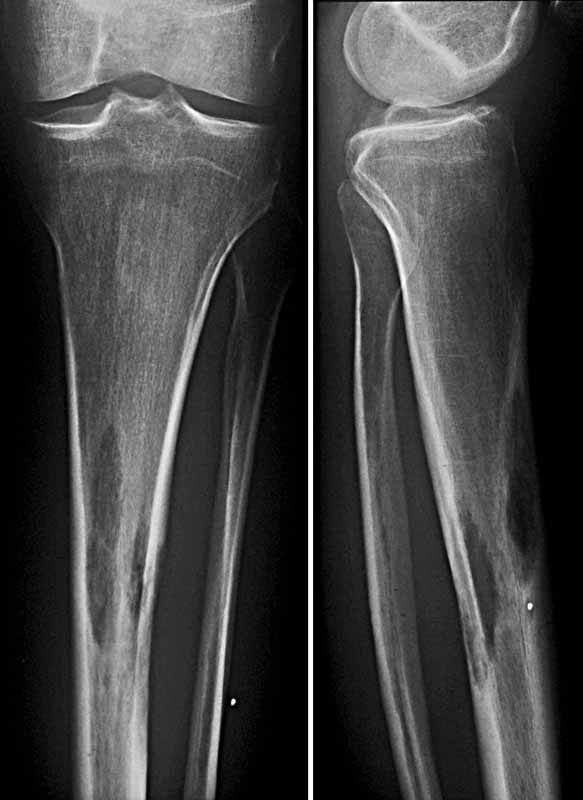
Bone metastasis of a bronchial carcinoma in the tibia.

Stardate: 92012.11

Today was my spinal tutorial session with my lecturer practitioner/clinical tutor, which ended up turning into a general tutorial session, giving us the opportunity to ask questions on anything we need help with. In the first half we took some x-rays, mainly lumbar spines and pelvic examinations. In the second half, we talked about image commenting, which was one of our final exams, and also makes up part of our clinical portfolio.

Basically, we take an image, any image, and describe what we see. Easy, right? Well, not always, but the process seems simple enough, which goes a little something like this:
  • Check the patient's clinical details - how old are they? What are their symptoms? 
  • Is the image, abnormal, possibly abnormal or normal?
  • Examine the general appearance of the bone
  • Trace the bone's cortex and density for any irregularities
  • Has the trabecular pattern been disturbed?
  • Give a detailed account of the abnormality seen
  • Examine the joint spaces and soft tissue
  • Are there any artefacts on the image?
I'll use the above image as an example, although I don't know the clinical details. Let's say the patient is a 29 year-old male, with severe pain around the tibia, for 3 months.
- The above image is definitely abnormal.
- The bone's cortex has not been disrupted, and the bone density appears normal. The trabecular pattern has not been disrupted.
- There are two radiolucent (the dark patch within the bone) area on the proximal end of the tibial shaft, visible on both projections, on the posterior and anterior tibial aspects.
- The joint space appears within normal limits, and the there is no soft tissue swelling
- There are no artefacts on the image.

This is a basic "comment", and as I found out, this image displays bone metastases.

Bone metastases, is a class of cancer metastases that is the result of primary tumour invasion of bone. Metastasis is the spread of cancer from one organ or part to another. Bone-originating cancers like osteochondroma for example, are rare. These metastases form solid masses, and bone is one of the most common metastasis location. 

Bone metastases can cause severe pain, bone fractures, spinal cord compression, and other major clinical concerns. These symptoms are caused by:
  • acidosis - increased acidity. Osteoclasts (bone cells that reabsorb bone tissue) generate extracellular protons, which lower the pH level.
  • bone restructuring - the uncoupled regulation of osteoclasts and osteoblasts (bone cells that form bone tissue) leads to bone malformation. Malformed bones are unable to withstand normal day-to-day mechanical stresses (e.g. weight bearing), leading to fractures, spinal cord compression and instability.
Anyway, this is post is getting long enough, and I've got dinner to make for myself. I hope this post has given you some insight into how we "examine" an image, in it's most basic form, and it's actually one of the fun parts of being a radiographer. True, there is a lot of terminology to get your head around, but once you have, everything sort of clicks into place! Tomorrow's my half-day and I'm once more in general, so hopefully I'll get the rest of my unaided examinations signed off.

LLAP guys!

References
  1. Bone Metastasis of a Bronchial Carcinoma in Tibia, n.d. photograph, viewed 29 May 2014 <http://www.mevis-research.de/~hhj/Lunge/imabc/BCKnMetb41_2.JPG>.

Wednesday, 28 May 2014

The 11-Week Stint

"How was your first day back in clinical?" "Great!"

Stardate: 92009.75

Heads up, True Believers! It's that time of term again.... placement! Bear in mind, this time round I'm here for 11 weeks, which followed on from our first year final exams (which I think went well, despite my original fears). What's that you say? I can relax now? Don't be ridiculous! I still have my portfolio, clinical assessments, an ALPS form, commenting forms, critique forms and a case study to produce yet! Does it make me miss Physics and Chemistry yet? A little...

This week (well, 2 and a half days), I'm placed in the General department, so I'll hopefully be seeing a good variety of examinations, get plenty signed off, and maybe, just maybe, get a chest assessment out of the way. It was strange going back into a clinical setting after being out for a while, especially seeing as I've been focusing so hard on revising for my exams. 

This morning happened to be a rather clunky one, (getting the 7:28 bus is too early), having stayed up late to iron all of my uniforms to save time. Getting ready with your eyes shut is never a good idea, by the way, even with my glasses on (there's a reason I keep my hair so short!) It also turns out, I forgot my placement notebook back in Leeds, which is what I attach my markers to (now jazzed up with Spider-Man stickers and polka dot patterns), so getting your images to the optimum standard, just isn't as easy. There's always something....

Once again, I felt like I'd forgotten everything, but after the first three pelvis x-rays, I was fine, considering the rest of my day consisted of... pelvis x-rays! Tomorrow is set to be my spinal tutorial, so I'll be practising spinal examinations (my favourite kind!) and critique. Finally, Friday is my half-day, so I'll be returning to Leeds for the weekend. A student radiographer's life is always busy!

I actually forgot how tiring placement is, no matter how much sleep you attempt to get beforehand. I got on the bus home and fell asleep! Luckily, I have the amazing knack to wake up just before my stop, and get off at the right one, so at least that's something. After forcing myself to make a healthy dinner, I traipsed into my room, I wrote this post, which will now be followed by a movie, a book and then bed!

Watch this space, it may get interesting...

Thursday, 1 May 2014

The Finals Countdown!

"So Emily, do you feel you're ready for your final exams?"

Stardate: 91935.01

Well, I'm back in lectures again for two weeks, after returning from my short Easter break (I'd say holiday, but as I was mostly revising and working, I'll refrain from doing so!) My first-year final exams are now looming ever closer, and despite revising every day, passing all the mock exams, and even printing off an A3 skeleton, I still don't feel ready. 

We've only got four exams, the longest of which being Physics at two and a half hours, but luckily they're spread out over four days, so I don't have to cram for two subjects at once beforehand. We've got two exams for Musculoskeletal Anatomy, one for Imaging Technique and one for Physics. I'm feeling confident for the Anatomy exam, as this is the one that, for now, makes the most sense. Physics on the other hand (see above image), well... writing pages and pages of notes has helped, but as far as 12-pulse bridge circuitry and rectifiers are concerned, I'm done for! You cannae change the laws of physics Jim, unfortunately...

Thankfully, a good chunk of some exams are MCQs (Multiple Choice Questions), and as long as you've done some reading around and understood the basics of the module, you should get most of these right, which usually can get you to the 40% pass boundary. Following those are the long answer questions, which range from 10-20 marks. 10 mark questions apparently only need about a page of writing, and 20 is two pages (...Wha?) So, my aim is to write as much as possible (within the relevant parameters anyway), and hope for the best!

After the exams are done with, we're back off out on placement again, but this time it'll be for 11 weeks! How will I survive 11 weeks of no Taekwon-do, nor a constant internet connection?! Do I risk taking my PS3, or shall I take boxsets upon boxsets of DVDs? Oh, decisions, decisions...

Wednesday, 9 April 2014

Portfolio Pains

What it feels like when you finish an examination for your portfolio and you try to get the radiographer to sign it.

Stardate: 91875.79

So, I'm in my last week of placement for this block, and this week is my second one in clinical skills. This is basically a week where you can take on real examinations, just a little slower, and focus on assessment techniques, as you are with a lecturer practitioner, and not on a busy department. It's actually a really good opportunity to get examinations signed off for your portfolio, and brush up on what you know, which catches up on you really quickly.

It also allows you to work with students from other placement sites and see the variations, as every Trust has their own Local Rules and protocols regarding examinations and safety etc. For example, where I'm placed, we may do different routine projections for an examination to another site, or technique may vary. Again, it all depends on where you're placed, and what the radiographers there have shown you.

The realisation has pretty much finally dawned on me that being a student radiographer is actually really hard! Balancing 7 weeks of holiday, placements, lectures, travel and expenses claims, revision, portfolios "social life", hobbies and sleeping into one academic year is not an easy task. In fact, it's knackering. Especially for students like me, who are crazy enough to have a part-time job too!

Getting portfolios filled in can be difficult for some sections, especially ours, as where I'm placed, certain types of examinations are few and far between, e.g. spines. Sure, you see plenty of chests, knees etc, but when a spine comes in, everyone wants to do it! Luckily, I'm almost done with signing off my portfolio, I just need a few more unaided examinations, then I'll take on the assessment run! (Better not leave it until the last minute.)

It's amazing how quickly a year goes by, as although it feels like September was ages ago, it also feels like it was yesterday I was standing in General X-Ray getting in everyone's way! We've got two weeks of holiday for Easter (I don't have a clue what I'm going to do for two completely free weeks!), then two weeks of lectures, an assessment week, and then all placement weeks until Summer!

I've got two more days to sign off as many things as I can, then I can relax for once (with revision sittings of course). If I can find the time, I may even go and watch the new 'Captain America: Winter Solider'!

Well, as I like to say... Geronimo and LLAP!

References
  1. The Hobbit, n.d. gif, viewed 9 April 2014,<http://31.media.tumblr.com/60b475d6d7c7ece10f1cda53b21de510/tumblr_ms3tfrXwoW1r9hlt2o8_250.gif>.

Saturday, 22 March 2014

A Week in Fluoroscopy

This is the equipment used in my placement hospital, the Siemens Artis Zee

Stardate: 91823.58

It's that time again: placement! And for week one, I was placed in Screening (AKA Fluoroscopy); I was supposed to be in A&E, but it was changed at the last minute. If you didn't already know what fluoroscopy is, it's basically an imaging technique that uses real-time moving images of the internal structures.

The equipment I got the chance to learn to use properly was the Siemens Artis Zee Multi-Purpose, and it's controls, although they look confusing, take very little time to get used to. It's used in various types of fluoroscopic procedures:
  • Root nerve blocks - an injection that is given close to a nerve as it leaves the spine; usually for sciatica pain.
  • Facet joint injections - an injection of local anaesthetic or steroid which can anaesthetise the facet joint to block pain. Facet joints are small joints between each segment of the spine.
  • Barium enema - an x-ray examination of the large intestine to help diagnose any existing problems affecting that area. The colon is filled with a contrast material containing barium, which is done by pouring the contrast into the rectum.
  • Barium swallow - an x-ray examination of the upper gastrointestinal tract, including the oesophagus, and sometimes the stomach. The patient is asked to drink a suspension of barium sulphate, and the x-ray images are taken during the swallow. This procedure is done in various positions: AP, oblique and lateral, for a 3D assessment.
  • Myelogram - an x-ray examination that uses an injected contrast material to view the fluid-filled space (subarachnoid space) between the spinal vertebrae. 
I witnessed these examinations, and there are more that are carried out on the department. I certainly got used to wearing lead rubber aprons, and quickly learnt to check the apron first, as instead of picking up the lighter 0.25mm apron, I picked up a heavy 0.35mm apron, which made me feel faint after a while! I also started using the thyroid shield, just to be on the safe side.

Despite what everyone first believes about barium enemas, you barely see (or smell) anything, and nearly all fluoroscopic procedures are painless for the patient (the barium mixture patients have to swallow has a pretty nasty after-taste, mind). 

Overall, it was nice to see what the department does, and what this technique involves. It certainly lets you know if you want to go into that sort of speciality, and it does actually seem quite interesting. You certainly see a good variety of patients, too. It's made me start seriously looking into what I want to do post-graduation (still definitely Forensic Radiography) as there are so many options with Radiography!

Until next time guys! Next week I'm in Clinical Skills, so we'll see how that goes.

LLAP!

References

  1. Siemens Artis Zee Multi-Purpose, n.d. photograph, viewed 22 March 2014 <http://usa.healthcare.siemens.com/siemens_hwem-hwem_ssxa_websites-context-root/wcm/idc/groups/public/@global/@imaging/@angio/documents/image/mdaw/mtc0/~edisp/artis_zee_multi_purpose-00068177/~renditions/artis_zee_multi_purpose-00068177~10.jpg>.

Tuesday, 11 March 2014

Mayhem in March

When people ask you about mock exams; applies to both preparation and execution!

Stardate: 91796.42

As you may have noticed, it's been quite a while since I last posted (and it has been ages, because I was on placement last time!) This is generally down to mock exams having been placed over the last two weeks. We had our exams in Musculoskeletal Anatomy, Medical Imaging Science, and Diagnostic Imaging Technique. 

I would like to say I was happy with my results, and I will, but I'm a picky human being with a funny little brain, and I know I definitely could have done better. So I'm happy, but not satisfied with them (to be fair, I haven't had one set of results back yet). I was surprised when I concluded that I found the physics exam the least terrifying!

They weren't so terrible in retrospect, but the only thing to remember about radiography exams is: write as much as you can that's relevant to the question! I lost marks for assuming I didn't need to write something down. If you've learnt it, write it down and talk about it! Also another handy tip is to definitely keep on top of your revision. It soon builds up if you don't!

Anyway, since I've managed to free myself from revision, as a 'reward', I've been reading some radiography journals to pass some time (when I'm not watching Star Trek, or Doctor Who). Two articles caught my eye in last month's (Feb 2014) issue of the journal Radiography. These were very different in content, but interesting reads.

The first was about using cross-sectional imaging in virtual post-mortem examinations. This appealed to me, generally because it's to do with Forensic Radiography, which is the field in which I want to continue my studies. The second was about professionals' experiences of imaging in the radiography process. I may have mentioned it before, but if you haven't already, signing up to the SoR is very beneficial for (prospective) radiography students, as you can access journals and articles, and loads of information which can give a nice scope of the field.

Soon you'll be hearing more from me again, as I'm back on placement next week! I'll be in A&E for one week, clinical skills for two, and a screening room for another. So there'll be plenty to get on with, and hopefully I'll have interesting things to feed back!

LLAP and reverse the polarity of the neutron flow!

References
  1. Matt Smith Filming His Final Scene, n.d. photograph, viewed 11 March 2014 <http://static.fjcdn.com/comments/Matt+Smith+after+filming+his+final+scene.+Leaving+the+TARDIS...for+_76a0889eed211250bf3e27e7c154b0c1.jpg>.