Thursday 17 September 2015

What Do You Mean I'm a Third Year?!

Definitely how I feel when it comes to university work!

Stardate: 93312.32

I realise I have been very non-existant on this blog as of late, but I've had a lot going on, and I completely forgot to write a post. After my mammography placement block, it was just a matter of getting my assessments completed and filling in our portfolio examinations, so I ended up returning to CT and similar departments. Following that, I had one exam resit (yay for reduced Summer holiday time) and lots of part-time work shifts. Oh, I also visited Dubai, too.

Now the realisation that I only have 10 months left until I graduate has finally dawned on me... I am terrified! That's barely any time, and soon enough, I'll be applying for jobs. I have suddenly lost the ability to do remember anything about x-rays... *sobs quietly in a corner*.

This year is when we have optional modules, as well as the compulsory ones:
  • Our Research Project (AKA the dreaded dissertation)
  • Professional Practice (the placement module)
  • Diagnostic Imaging Technique
  • Pathophysiology
  • Preparation for Practice
  • 2 optional modules, of which I chose a Focused Practice module and Advanced Medical Imaging Systems
  •  and finally the elective weeks in June.
I have some idea on what my dissertation will be, but I'll try to avoid thinking about it again until it's mentioned in a lecture... just to be on the safe side. Especially after looking at how many essays we have to hand in this year, the above gif works wonderfully. This year is going to be... hard. As for my electives, I've been playing with the idea of going abroad, or spending some time in a CT department... as I can't do anything Forensic related until I graduate. I'm not ready to be a full-blown adult yet...!

Either way, I apologise for the lack of posting, I will pick it back up as soon as the semester starts next week!

LLAP!

Thursday 25 June 2015

How To Not Be Awkward in Mammography Departments

Fact: No-one enjoys having a mammogram. No-one!

Stardate: 93085.08

I've gone pink for this post, (I know it's not October, but for this post I wanted to give a quick nod to the Breast Cancer Awareness organisation, Pink Ribbon) so if you have trouble reading it, I do apologise! I have also dedicated this post to my amazing and brave Auntie Mich, who kept me motivated to keep this blog going way back when I was just starting out!

It was the one placement I was openly dreading. After already having done radiography for nearly two years, you'd think I was already okay with seeing humans undressed in most ways, shapes and forms. No matter how many times you tell patients they can keep their underwear on, there's always one who didn't hear you... Apparently, I was not fully prepared.

I have to admit, despite having heard and been taught about Mammography, I'd never taken an active interest in the modality, nor fully looked into it. But once I'd looked into (aka Googled) the basic physics of mammography equipment  to refresh my memory post-exams (e.g. ultrasound, mammography units), I started to warm more to the subject than I had previously. The other plus side post-graduation wise: men currently can't do the job, so there is less competition for job applications (although I don't agree with this, from a work gender-equality angle).
Mammography is basically low energy x-rays that image the breasts. It's most well-known for its being an early detector of breast cancer, by finding masses or microcalcifications. I spent time in both screening clinics, where women who fit the criteria are checked every few years for any changes; and assessment clinics, where women have attended their GP after having found a lump, for example. 

So last Monday after having arrived at the department and been shown around, I witnessed my first mammogram, and couldn't help but look everywhere in the room, except at the body parts being examined. Eventually, I had to buck up and pay attention, or last week's block was going to go pretty slowly for me.

Every patient I saw examined was happy for me to stand in the room and have everything explained to me, and some even explained instead of the mammographer, which was a new experience! But this did put me more at ease, as being a student I'm used to doing the 'Student Shuffle' (basically constantly moving out of everyone's way) and being quiet when not in the general radiography or CT departments.

Once I'd followed a few women through from their mammograms to their ultrasound, I started to gain more interest and look in more detail at the images. I particularly enjoyed helping one of the consultant radiographers (yes, you read that correctly, we can become consultants!) with an ultrasound guided biopsy, where I was shown different types of tissue structures within the breast that would be going for testing.

I also got to spend one morning sitting with a doctor while he ran a clinic. It was interesting to see the other side, watching the pre-assessment, and then seeing how the imaging side was requested and for what reasons.

However, with Mammography, you will eventually come across sad news at some time or another. A number of the patients I saw examined at the department had already had a mastectomy (breast tissue removal surgery), and some images I was shown for comparison and to help me understand breast anatomy, displayed what were cancerous masses. For me, this was quite a tough experience (hence why I've never considered Radiotherapy), and it hammered home how beneficial Mammography is as a department. Even for a small number of men.

From what I've witnessed, being a mammographer, is a very intimate job. During the examination, you will be in the patient's personal space, and the procedure can be difficult and uncomfortable for patients. It's a difficult job to do, putting apprehensive and worried women at ease, and if they're upset, you have to be the type of person who knows how to handle that situation kindly and professionally. Not only that, nothing can be missed off the image, because something small could be hiding in that one area not included, so you have to be skilled enough to make sure you've gotten everything on the receptor. There is also a chance to be a breast sonographer, giving you a little more autonomy in your work.

Personally, I'm not completely sold on Mammography myself. If I had to say what I enjoyed the most, it would be the ultrasound examinations and biopsies, as I could 'see' more on those images, than on the plain film ones (a slightly odd situation for an undergrad radiographer). I wouldn't mind going back for another week, but right now, it's not something I could see myself going towards once I graduate. I can see why it's an appealing job to some, and don't get me wrong, I found the diagnostic side of it and the variation interesting, but all day and every day would be breasts. And I don't think I'm ready for that just yet!

This week I'm in the Paediatric department witnessing all sorts of examinations. But, more on that in my next post!

LLAP Guys!

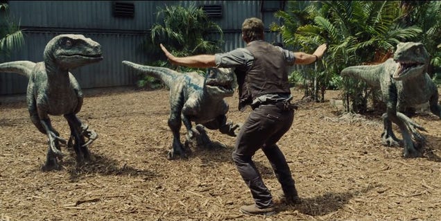
Ps. I hope you've all gone to see Jurassic World... 

Thursday 18 June 2015

Managing Placement Assessments, and CT in (Dino)Nutshell!

How I feel handling all my different placements and assessments...

Stardate: 93066.03

I apologise, as this post is a few (ahem... five) days late. This is once again because of part-time work, and placement in a different hospital due to the department I'm in this week (more on that in my next post!)

As I'm now in my "Summer stretch" of placement blocks, second year clinical assessments are now creeping up on me, rather ominously too, as they're not easy to pick appropriate patients for! And as you have so little time in each department, due to rotations, every moment there is precious. No-one ever said second year was easy... Including me!

So far, I've passed my CT head and Extradepartmental Chest X-ray (Mobile/Portabe) assessments; so only three more to go! To celebrate passing my CT assessment, I decided to dedicate this post (briefly) on CT, and also went to see 'Jurassic World'... my inner child was incredibly happy (so was the grown-up, because Chris Pratt; 'nuff said.) It's amazing, I highly advise everyone to see it.

So, about CT, or Computed Tomography! Most people have heard of, or at least have had, a CT (or CAT) scan. For Leeds university students, CT is a department we visit, to further our understanding of extradepartmental modalities, that involve medical imaging. CT scans, use a 360-degree x-ray beam to create a 3D image, using a number of imaged slices taken from a large series of two-dimensional images, to visualise the internal structures of the body, without having to cut open the patient! CT has plenty of applications, including the detection of abnormalities, as well as diseases, such as cancer, inflammation and trauma etc.

There are a multitude of advantages to CT over the usual 2D medical radiography. It doesn't have the issue of superimposition (organs/tissues lying over each other), allowing structures to be assessed, and allowing the area of interest to be studied in detail. CT also has a high-contrast resolution, meaning that tissues with even the slighest difference in density can be distinguished from one another. Finally, CT images can be viewed in various ways, or "planes", allowing any exisiting pathology's position to be pin-pointed.

Non-medical uses of CT include discovering flaws in industrial parts before they are used, and to identify the existence of any oil, water or gas etc. in structures.

I find CT an interesting department to look into once I've qualified, as they're used quite often in forensic radiography for virtual autopsies, which I'm thinking of writing about for my final-year dissertation. Being able to see all the internal anatomy without it being superimposed onto itself is great, as it makes spotting abnormalities that little bit easier... but not by much for the inexperienced!

And that is basically CT in its barest form! This week I've been in the Mammography department, so I'll have a more in-depth post coming up regarding the Breast Screening process, and a little on the modified equipment.

Final Note: GO AND SEE 'JURASSIC WORLD'. Think I made that clear enough...

LLAP Guys!

Monday 1 June 2015

So It's Been 5 Months, Eh? Let Me Tell You About Cardiac Cath Labs...


How I feel looking in the mirror now I have to get up early for placement...

Stardate: 92967.34

Well, aren't I just the worst blog owner? But in all fairness to myself, I have been entirely swamped by being a student radiographer. My placement in January got incredibly hectic, I've worked so many shifts at my part-time job, and then there's these evil little things our lecturers call 'exams' that recently came and went... But, finally I'm back on placement, so I'm going to be right back on track with this blog. I mean... I'm sure you've all missed it, right?

Now, if any of you readers are prospective students or currently a first year at the University of Leeds, let me give you another bit of advice... Second year is freaking hard. I have never felt so mentally drained in my life! And I've worked as a full-time supervisor in retail...  

Don't get me wrong, I still love my university and the course, because I know it'll be worth it in the end. Our lecturers do an amazing job, but there is just so much to cram into your head, you'll be begging June to come faster so the May exams are over. And I thought the January exams were tough! Never have I been so wrong...

But it's not all doom and gloom! Hooray! 

So, what are Cardiac Cath Labs, you ask? Well, in the UK, a Cath(eter) Lab is an examination room containing diagnostic imaging equipment used to visualise the arteries and chambers of the heart for treatments or investigations. They're staffed by a multidisciplinary team such as cardiac consultants, cardiac physiologists, nurses and radiographers. 

Typical examinations that occur in Cath Labs include:
  • Coronary catheterisation
  • Coronary angioplasty or Percutaenous Coronary Interventions - used to treat stenotic (narrowed) coronary arteries
  • Transcatheter Aortic Valve Implantations - the insertion of an artificial heart valve using catheters
  • Pacemaker implantations
  • Electrophysiological studies - e.g. mapping of the heart to locate heart abnormalities
  • and Cardiac ablations - a procedure that is used to scar small areas in the heart that may be causing a heart rhythm problem.
I managed to see a good range of procedures this week, but the most interesting I saw was probably the PCIs, as radiographers have a large role in the team. PCIs usually use a femoral (leg) or radial (forearm) artery to access the coronary arteries of the heart. Then through a 'sheath', a guide catheter is pushed to the opening of the coronary artery; this allows contrast (x-ray dye) to be injected into the artery. The size of the artery is then assessed to select what size balloon catheter will be used. The guide catheter is moved to the stenosis (narrowing), so the balloon catheter can inflate, forcing any 'plaque' to compress, and the arterial walls to expand.

I also saw a TAVI which involved a large team, and plenty of contrast to check the valve's operation. I watched as the valve was prepped, and the one I was lucky enough to see was made from hand-sewn strips, and formed from pig pericardium (a double-walled sac that contains the heart)! It's amazing how small the valves become before they're inserted, and then you get to see them deploy to their full size around the old valve site. Seeing the valve outside the body made me appreciate how much magnification is used in fluoroscopic imaging!

Unfortunately, there isn't too much I can say, as I only went to Cath Labs for 3 days, and it went by so quickly! And as a radiography student, we can only do so much in that department with our basic training. It seems I need to get used to the early mornings and placement again!

Next week I'm working the Late shifts over the weekend, but this time, not as a radiographer assistant, but as a student. So I'll probably have plenty to talk about!

Once again, sorry for the long delay, but I'm hoping to keep on track once more!

LLAP!

Monday 26 January 2015

Ultrasound - Not Just Babies!

  An ultrasound scan for the thyroid gland.

Stardate: 92672.31

Last week saw me in the hospital's various ultrasound departments: general, obstetric, paediatric and vascular. Despite my initial reservations and disinterest in ultrasound, I actually really enjoyed my week there. At first, I tried to read up on basic anatomical appearances, and how ultrasound images were produced, including the physics and which transducers (probes) were used for which examinations etc. But I'm going to leave that for another post... 

From my basic understanding, I started to get the hang of it all, and could get my head around what position the transducer was in when I observed examinations. I even got to have a go in some departments, where I managed to scan a kidney, my own thyroid gland, and finally an abdominal aortic aneurysm (AAA).

In the general department, the examinations are mainly musculoskeletal and some gynaecological procedures. Obstetric is the most famous ultrasound department, as it's where a multitude of women go for their pregnancy dating, 12-week and 20-week scans. Usually the 20-week scan is an anatomy check, as well as to check the sex of the baby (if the parents want to know what they're having). Paediatric is of course the children's department of ultrasound. And finally, Vascular is where all the circulatory system investigations occur.

Anatomy on ultrasound scans takes a little bit longer to get your head around than general x-ray anatomy, but once you've witnessed a few scans, you start to recognise certain structures and their appearances. If I do take anything away with me from my week, it is that fluid always look black and veins, in comparison to arteries, are compressible. Which is how sonographers test for DVT (Deep Vein Thrombosis).

As ultrasound has such a wide range of examinations and procedures, I couldn't really pinpoint one I wanted to talk about in detail that would fit into this post nicely, so I'll possibly put one up soon after some serious consideration! There were plenty I witnessed where I wouldn't mind looking further into them.

Out of all the departments I visited, the most interesting I found to be was Vascular, but each department has its own intrigue. Being placed in ultrasound has certainly changed my attitude towards the modality, and I actually wouldn't mind going there again. Some students find it boring, but if you ask plenty of questions and try to look into image production and anatomy before you go, there's less chance you'll be confused, and will be able to identify what you're actually looking at!

This week I'm in A&E, but I'm working alongside the first-years, so now I have to look like I actually know what I'm doing! I've also started my radiography assistant job, so you may start seeing posts about doing part-time work alongside the course cropping up!

LLAP guys!

References
  1. Diagnostic Ultrasound Scan, n.d. photograph, viewed 25 January 2015 <http://www.ultrasoundnow.co.uk/wp-content/uploads/2012/01/shutterstock_82853839.jpg>.

Friday 16 January 2015

The January Exams Episode

January exams are over it seems...

Stardate: 92647

I know, I know, it's been forever since I made a post... well, nearly two months anyway, if I'm not mistaken. But I do have an entirely reasonable excuse why. During this last week, I have been studying like a demon for my three January exams that snuck up on me over the Christmas period. Apparently, being a second year is hard. No, I mean it. Really hard.

We had three exams, one for Research Stats (this module, ladies and gentlemen, I kid you not, is pure evil. Sauron himself probably would have agreed), Chest Commenting and Anatomy. No matter how much I tried to get my head around Research Stats, it would not sink in, and I have the horrible, foreboding feeling that I've failed the exam. Commenting by far was the exam I was most confident for, and Anatomy I tried to write loads and draw as many diagrams as I possibly could! All that, and I also had a Physics essay to hand in at the start of the assessment week. So, with all that, I'm knackered.

But now that it's all over, I get a nice weekend to myself, (even after doing a quick shift in my new job as an radiography assistant - woo!) where I don't have to think about anything. That is until Monday rolls around, and the early mornings begin again... you guessed it, placement is here again!

This time around is a 5-week block. I'm in ultrasound, A&E, CT, fluoroscopy and finally orthopaedics. So yes, I'm going to be nice and busy. I'm looking forward mainly to CT, as it'll be a new experience for me. But I'm having to start thinking about the dreaded assessments, which will be:
  • GI Tract with contrast
  • Circulatory/Renal/fluoroscopy examination with contrast
  • MSK non-ambulant
  • Extra-departmental chest x-ray (e.g. a portable)
  • CT Head without contrast 
We'll see how it all turns out. But at least ultrasound will be slightly more relaxed than some other departments anyway. Hopefully they're nice to me on my birthday this Wednesday...

I'd better start looking at how ultrasound actually works...

LLAP!