Monday 5 November 2012

Lack of Posting Apology


Hello everyone! I am writing this blog entry, due to the fact I have noticed how long ago it was that I wrote an entry here. The reasons behind this lag is due to both work, college and extracurricular busyness. I apologise for my silence, but when I eventually get back around to typing entries they will be back to my normal standard.

To keep you updated on how I'm doing, my personal statement is finished, I just need to have someone check it over, and my university application is complete! 

Good luck to all those reading this who are going through the same process!

Monday 22 October 2012

WOTD: Onychogryphosis

The effects of onychogryphosis. Photograph: Elord @wikidoc.org

"Onychogryphosis" - Abnormal thickening, hardening, and curving of the nails that occurs mainly in elderly people. Onychogryphosis may be associated with fungal infection or poor circulation.

"onychogryphosis". British Medical Association (2007). Illustrated Medical Dictionary. 2nd ed. London: Dorling Kindersley (DK). p414.

Saturday 20 October 2012

X-Ray Generators

An interventional radiology theatre. Photograph: bsir.org

(Hey guys, sorry I haven't posted in a while!)

X-ray generators are devices that are used be radiographers to acquire images that are taken using x-rays to see the body's internal structures. They can also be used in sterilisation and fluorescence scans.

The main component of the x-ray generator is the x-ray tube. This tube contains a cathode which directs an electron-stream (don't cross the streams!) into a vacuum, and an anode which collects the electrons. The anode is made from tungsten, molybdenum, or copper, and when the electrons collide with it, only about 1% of the resultant energy is emitted as x-rays.

Many scientists have been involved with the development of x-rays, such as Hermann von Helmholtz, Johann HittorfWilliam Crookes, Heinrich Hertz, Nikola Tesla, and of course Wilhelm Röntgen

X-ray imaging systems generally consist of an x-ray source, an image detection system, and a PACS. These imaging systems are used for many different applications, the most common and well-known one being for health-care/medicine

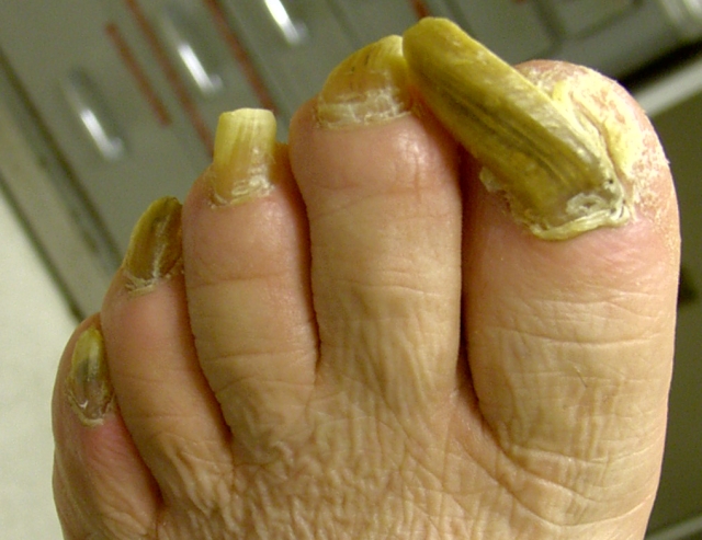
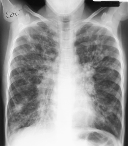
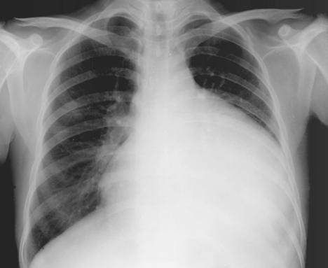
In medicine, the two main fields x-rays are used is radiography and fluoroscopy. Radiography is used to produce fast images of the internal bone structures and tumours, as well as treating them (radiotherapy). Fluoroscopy is used when real-time imaging is necessary. These types of x-rays are highly penetrating, ionising radiation and therefore are only used on dense tissues, like bones, as they absorb the radiation better than soft tissues. 

X-ray machines are also used in security measures. The most common of these is in airports or in schools. These x-rays are used to examine luggage for weapons, bombs. These x-ray systems are expensive but are non-invasive.

An advance in x-ray technology recently has been to the cathode which can be made from carbon nanotubes. These have been shown to provide 3-D images in less time than conventional x-ray machines do. Also, as they use less energy, they accumulate lower operational costs.

Saturday 13 October 2012

WOTD: Prolactinoma

Side-view MRI scan showing a Prolactinoma. Photograph: UCLA Neurosurgery

"Prolactinoma" - A noncancerous tumour of the pituitary gland the causes over-production of prolactin. In women, this may result in galactorrhoea, amenorrhoea, or infertility. In men, it may cause erectile dysfunction and gynaecomastia. In either sex, it may cause headaches, diabetes insipidus, and, if the tumour presses on the optic nerves, loss of the outer visual field. Diagnosis is made from blood tests and CT scanning or MRI of the brain. Treatment may involve removal of the tumour, radiotherapy or giving the drug bromocriptine.

"prolactinoma". British Medical Association (2007). Illustrated Medical Dictionary. 2nd ed. London: Dorling Kindersley (DK). p466.

Thursday 11 October 2012

WOTD: Embolism

A CT scan showing a pulmonary embolism. Photograph: mediexchange.com

"Embolism" - Blockage of an atery by an embolus. Blood clots that have broken off from a larger clot located elsewhere in the circulation are the most common type of embolus. Pulmonary embolism is usually the result of a fragment breaking off from a deep vein thrombosis and being carried via the heart to block an artery supplying the lungs; this is a common cause of sudden death. Blood clots may form on the heart lining after a myocardial infarction, or in the atria in atrial fibrillation, and then travel to the brain, resulting in a cerebral embolism, which is an important cause of stroke.

Symptoms of an embolism depend on the site of the embolus. Pulmonary embolism can lead to breathlessness and chest pains. If the embolus lodges in the brain, a stroke may occur, affecting speech, vision, or movement. If an embolism blocks an artery to the leg, the limb will become painful and turn white and blue. Untreated, gangrene may develop. In serious cases of fat embolism, heart and breathing rates rise dramatically, and there is restlessness, confusion, and drowsiness.

Embolectomy (surgery to remove the blockage) may be possible. If surgery is not possible, thrombolytic and anticoagulant drugs may be given.

"embolism". British Medical Association (2007). Illustrated Medical Dictionary. 2nd ed. London: Dorling Kindersley (DK). p196.

Tuesday 9 October 2012

Radiography Assistants & Imaging Support Workers

A CT scanner team consists of two radiographers, a radiologist and a radiography assistant.
Photograph: NHS

Okay, so I was looking into my options if the unexpected happened e.g. if I  :
a) didn't pass my Access course this year
or
b) didn't manage to get a place onto a university course. And I was looking around, and I discovered support roles in radiography! (Even if I don't get a place for 2013, I will still apply again for 2014!)

Radiography assistants and imaging support workers are people who assist qualified diagnostic/therapeutic radiographers. These assistants have certain responsibilities depending on the nature of their team's work.

They generally aid patients as part of their job role, but their other duties include:
  • processing film images
  • preparation and assistance in general/specialised procedures 
  • keeping the working environment hygienic and clean
  • preparing treatments
  • manual handling of patients
  • basic equipment maintenance
  • recognising and reporting faulty equipment
To work as a radiography assistant/imaging support worker, there are no formal entry requirements, but 4 or more GCSEs, or their equivalent may be required for more advanced work. There is an NVQ available, but it is not a route towards qualifying as a radiographer. It is also possible to get into this role via a level 3 apprenticeship.

These workers are usually train while on the job, but there are short courses available. The Society of Radiographers (SoR) has developed accreditations for assistant practitioners working in these settings. It is to ensure the qualifications and training is recognised by the NHS. It is also possible to work while studying part-time towards a professional radiography degree.

So, again, there's always a way to learn about the job even if the scary things happens!

WOTD: Venepuncture

A contrast-enhanced venography in a 78 year-old man with thrombosis of the deep veins in the lower leg.
Photograph: ajronline.org

"Venography" - A diagnostic procedure that enables veins to be seen on an X-ray film after they have been injected with a substance opaque to X-rays. It is used to detect abnormalities or diseases of the veins, such as narrowing or blockage from thrombosis.

"venography". British Medical Association (2007). Illustrated Medical Dictionary. 2nd ed. London: Dorling Kindersley (DK). p581.

Saturday 6 October 2012

WOTD: Barotrauma

Chest x-ray of Barotrauma in a patient with status asthmaticus. Photograph: realicu.com

"Barotrauma" - Damage or pain, mainly affecting the middle ear and facial sinuses, that is caused by changes in surrounding air pressure. Air travellers are at the greatest risk, but scuba divers face similar problems.

Aircraft cabin pressure decreases as the plane ascends and increases as it descends. As the aircraft ascends, the ears may "pop" as the air in the middle ear expands and is expelled via the eustachian tubes, which connect the middle ear to the back of the throat. On descent, the higher pressure may push the eardrum inwards and cause pain. Minor pressure damage in the middle ear may cause pain, hearing loss, and tinnitus for a few days; damage within the facial sinuses may also cause pain, and possibly a discharge of mucus or blood. Symptoms usually wear off within hours or days, but treatment may be needed if they worsen or persist. Large pressure changes can rupture the eardrum.

Barotrauma can be avoided by vigorous swallowing or by forcibly breathing out with the mouth closed, and the nose pinched (the Valsalva manoeuvre). This action equalises the internal and external pressures in the middle ear and sinuses. If the eustachian tubes are blocked, as commonly occurs with a cold, use of a nasal spray containing a decongestant drug is recommended shortly before the descent of the aircraft. 

"barotrauma". British Medical Association (2007). Illustrated Medical Dictionary. 2nd ed. London: Dorling Kindersley (DK). p66.

Tuesday 2 October 2012

CT and Sir Godfrey Hounsfield

Sir Godfrey Newbold Hounsfield. Photograph: mc.vanderbilt.edu

Sir Godfrey Newbold Hounsfield CBE, FRS (28 August 1919 - 12 August 2004) was an English electrical engineer who shared the Nobel Prize for Physiology or Medicine with Allan McLeod Cormack, for his part in developing the diagnostic technique of X-ray computed tomography

The Hounsfield scale, a measure of radiodensity used in CT scan evaluations, is named after him. The scale's units is defined in Hounsfield Units.

Godfrey was born in Sutton-on-Trent, England. He had two elder brothers and two elder sisters. He was intrigued by electrical gadgets, and he tinkered with his own electronic machines, launched himself off haystacks with his own home-made glider, and almost killed himself by using water-filled tar barrels. He went to Magnus Grammar School and excelled in physics and mathematics.

Just before World War II, he joined the Royal Air Force, where he learned the basics of electronics and radar. After, he attended Faraday House Electrical Engineering School. He graduated with a Diploma of Faraday House.

In 1951, he began working at EMI Ltd, where he began researching guided weapon systems and radar. It is here he became interested in computers and helped design the first commercially available all-transistor computer: the EMIDEC 1100. 

Hounsfield came up with the idea that one could determine what was inside a box by taking X-ray readings at all angles around the objectHe then constructed a computer that could take X-rays input at varying angles to create an image of the object in "slices". Applying this to the medical field led him to propose what is now known as computed tomography. At the time, Hounsfield was not aware of Cormack, who had done work on the theoretical mathematics for such a device.

Hounsfield built a prototype head scanner and first tested it on a preserved human brain, then on a fresh cow brain from a butcher shop, and later on himself. On 1 October 1971, CT scanning was introduced into medical practice with a successful scan on a cerebral cyst patient at Atkinson Morley Hospital, London. In 1975, Hounsfield built a whole-body scanner.

WOTD: Talipes

Left congenital talipes as seen by three-dimensional ultrasound at 22 weeks gestation. 
Photograph: International Federation of Gynecology and Obstetrics

"Talipes" - A birth defect (commonly called club-foot) in which the foot is twisted out of shape or position. The cause may be pressure on the feet from the mother's uterus, or a genetic factor. The most common form is an equinovarus deformity, in which the heel turns inwards and the rest of the foot bends down and inwards. It is treated by repeated manipulation of the foot and ankle, starting soon after birth. A plaster cast, or strapping may be used to hold the foot in position. If this is not successful, surgery will be needed.

"talipes". British Medical Association (2007). Illustrated Medical Dictionary. 2nd ed. London: Dorling Kindersley (DK). p538.

Monday 1 October 2012

WOTD: Retinoblastoma

A young child with retinoblastoma in the left eye. Photograph: Gordon K. Klintworth

"Retinoblastoma" - A cancer of the retina that affects infants. The first indications of this disorder may be a squint caused by blindness in the affected eye or a visible whiteness in the pupil. Without early treatment, retinoblastoma can spread to the orbit (eye socket) and along the optic nerve to the brain.

Retinoblastoma has a genetic basis. People with this cancer lack part of one of the chromosomes in pair number 13. Newborn infants from affected families should have genetic counselling.

Treatment is by removing the eye, or by radiotherapy. If both eyes are involved, the one worse affected by removed and the other given radiotherapy.

"retinoblastoma". British Medical Association (2007). Illustrated Medical Dictionary. 2nd ed. London: Dorling Kindersley (DK). p489.

Sunday 30 September 2012

WOTD: Phaeochromocytoma

Phaeochromocytoma on an MRI scan. Photograph: endocrinediseases.org

"Phaeochromocytoma" - A rare tumour of cells that secrete epinephrene (adrenaline) and norepinephrine (noradrenaline). It causes increased production of these hormones, leading to hypertension. The tumours usually develop in the medulla (core) of the adrenal glands, and are most common in young to middle-aged adults.

Hypertension is the only sign most of the time, but pressure on the tumour, emotional upset, change in posture, or taking beta-blocker drugs can cause a surge of hormones. This surge brings on a sudden rise in blood pressure, palpitations, headache, nausea, vomiting, facial, flushing, sweating, and, sometimes, a feeling of impending death.

Blood tests and urinalysis are used to make a diagnosis, CT scanning, MRI, and radioisotope scanning may be used to locate the tumours, which are then usually removed surgically. Follow-up medical checks are required because the condition occasionally recurs.

"phaeochromocytoma". British Medical Association (2007). Illustrated Medical Dictionary. 2nd ed. London: Dorling Kindersley (DK). p445.

Saturday 29 September 2012

Fluoroscopy

An example of a fluoroscopy. Photograph: spineuniverse.com

Fluoroscopy is an imaging technique commonly used to obtain real-time moving images of the body's internal structures through a fluoroscope. In its simplest form, a fluoroscope consists of an x-ray source and a fluorescent screen. Modern fluoroscopes combine the screen with an x-ray image intensifier and a CCD video camera, which allows the images to be recorded and played on a monitor.

The use of the x-rays requires that the potential risks be balanced against the benefits the fluoroscope to the patient. Although low doses are always tried to be used during a fluoroscopic procedure, the general length of a typical procedure results in a high absorbed dose to the patient.

They can be used to:

  • investigate the gastrointestinal tract
  • aid fracture reduction and the placement of metalwork during orthopaedic surgery
  • carry out an angiography of the heart, leg and cerebral vessels
  • aid urological surgery

WOTD: Scoliosis

A scoliosis x-ray. Photograph: thefamilychiro.com.au

"Scoliosis" - A deformity in which the spine is bent to one side. The thoracic or lumbar regions are most commonly affected. Scoliosis usually starts in childhood or adolescence and becomes progressively more marked until growth stops. In many cases, another part of the spine curves to compensate, resulting in an S-shaped spine. The cause of juvenile scoliosis is unknown. Rarely, scoliosis is due to a congenital abnormality of the vertebrae.

In some cases, physiotherapy may be sufficient to control scoliosis. Progressive or severe scoliosis may require immobilisation of the spine in a brace, followed by surgery (spinal fusion) to straighten it.

"scoliosis". British Medical Association (2007). Illustrated Medical Dictionary. 2nd ed. London: Dorling Kindersley (DK). p500-501.

Friday 28 September 2012

WOTD: Bronchiectasis

A chest x-ray of a patient suffering cystic fibrosis with bronchiectasis. Photograh: learningradiology.com

"Bronchiectasis" - A lung disorder in which one or more bronchi (the air passages leading from the trachea) are abnormally widened, distorted, and have damaged linings. Bronchiectasis most often develops during childhood and was once commonly associated with infections such as measles and pertussis (whooping cough). The condition is also a complication of cystic fibrosis. It results in pockets of long-term infection which the airways and the continuous production of large volumes of green or yellow sputum (phlegm), Extensive bronchiectasis causes shortness of breath. The symptoms are usually controlled with antibiotic drugs and postural drainage. If the condition is confined to one area of the lung, surgical removal of the damaged area may be recommended. 

"bronchiectasis". British Medical Association (2007). Illustrated Medical Dictionary. 2nd ed. London: Dorling Kindersley (DK). p98.

Thursday 27 September 2012

Arthrograms

The injection of an iodine-based contrast medium arthrogram. Photograph: Zezounet

An arthrogram is a series of images (mainly x-rays) of a joint after it has been injected with a contrast medium. These injections are usually administered under local anaesthetic. It is performed by a radiologist who uses fluroscopy and ultrasound to guide the placement of the needle into the joint, to then inject the contrast fluid.

Shoulder arthrograms are used to study tears of the rotator cuff (mainly tears of four tendons rather than the muscles in the shoulder). They can also define whether there are abnormalities of the glenoid labrum and bicipital tendon

Arthrograms (like radiography) can either be diagnostic or therapeutic. Therapeutic arthrograms are often joint distension and cortisone injection procedures. This procedure is most common for the shoulder. Diagnostic arthrograms can be direct or indirect, by a venous injection of contrast material and then followed by a CT or MRI scan.

Arthrograms are not not recommended to patients who are allergic or sensitive to:
  • medications
  • contrast dyes
  • local anaesthesia
  • iodine
  • latex
Infection in the puncture site where the radiopaque substance is injected is a small risk, as is bleeding.

There have been reports that gadolinium contrast agents cause Neophrogenic Systemic Fibrosis (NSF). This has only occurred in cases where the patients have moderate-to-end-stage kidney disease; there have been no reports stating any case of NSF in those with healthy kidneys. Other than the risk of NSF for those enduring kidney disease, arthrograms carry the same risks as any ordinary X-ray scans.

WOTD: Spondylolysis

A sagittal x-ray of a female patient with L4 spondylolysis. Photograph: Spineuniverse.com

"Spondylolysis" - A disorder of the spine in which the arch of the 5th (or, rarely, the 4th) lumbar vertebra consists of soft fibrous tissue instead of normal bone. As a result, the arch is weak and prone to damage, which may produce spondylolisthesis (the slipping forward of a vertebra over the one below it.) Otherwise, spondylolysis is usually symptomless.

"spondylolysis". British Medical Association (2007). Illustrated Medical Dictionary. 2nd ed. London: Dorling Kindersley (DK). p522.

Wednesday 26 September 2012

John Wild: The Father of Ultrasound

John J. Wild. Photograph: Boston.com

John Julian Cuttance Wild (11 August 1914 - 18 September 2009) was an English-born American physician, who was part of the first group to use ultrasound for imaging the body, most notably for diagnosing cancer. The modern ultrasonic diagnostic medical scans are improvements upon the equipment Wild developed in the 1950s. He has since been referred to as the "Father of Medical Ultrasound".

Wild was born in Kent, United Kingdom in 1914. He attended Merchant Taylor's School and took out his first patent at the age of 14, for a device to distribute an even amount of hot and cold water in a bathtub. He received a Bachelor of Arts degree in Natural Sciences Tripos from the University of Cambridge in 1936. In 1940, he was awarded a Master of Arts. And he also became a Doctor of Medicine in 1942. He was elected a member of the Royal Society of Medicine in 1944 and then, in the same year joined the Royal Army Medical Corps

In 1946, he emigrated to the US and began working at the University of Minnesota in the department of surgery. In 1950, he switched to the electrical engineering department. During World War II he had seen many patients that had suffered with bowel failure and he had developed a technique called the "Wild Tube". In the US, he saw patients with the same conditions, and so he conceived the idea of using ultrasound as a non-invasive means of determining the intestinal injuries of patients. The sound bounced back from the tissue, identifying its thickness and resilience of the portion of intestine being scanned. He had developed his idea, after hearing of high-frequency sounds being used as a means to identify cracks in tank armour. 

The first machines did not have a strong enough resolution to scan the intestines, but by 1951 Wild and Dr. John Reid gained access to equipment that operated at 15 MHz, which providing the detail needed to carry out internal scans and differentiate between cancerous and healthy tissue. 

In 1991, Wild earned the Japan Prize, in recognition of his work within the field of ultrasound imaging

WOTD: Osteochondroma

An osteochondroma on the fibula. Photograph: tumorlibrary.com

"Osteochondroma" - A noncancerous bone tumour, which is formed from a stalk of bone capped with cartilage, and appears as a hard round swelling near a joint. An osteochondroma develops in late childhood and early adolescence, usually from the side of a long bone near the knee or shoulder. The tumour causes problems only if it interferes with movement of tendons or the surrounding joint, in which case it may be removed surgically. Large osteochondromas can interfere with skeletal growth, causing deformity.

"osteochondroma". British Medical Association (2007). Illustrated Medical Dictionary. 2nd ed. London: Dorling Kindersley (DK). p422.

Tuesday 25 September 2012

Paul Lauterbur & MRI

Paul Christian Lauterbur. Photograph: Michigan State University

I decided to look into the development of MRI today, and I learnt about somebody new!

Paul Christian Lauterbur (6 May 1929 - 27 March 2007) was an American chemist who shared the Nobel Prize in Physiology or Medicine in 2003 with Peter Mansfield for his work which made the development of MRI possible.

Lauterbur was born and raised in Sidney, Ohio and graduated from Sidney High School where a new wing for Chemistry, Physics and Biology was dedicated in honour of him. He studied as an undergraduate at Case Institute of Technology  in Cleveland. He built his own laboratory as a teenager in his parent's basement, and he enjoyed doing chemistry experiments on his own. While in the Army in the 1950s, he began his work developing one of the early MRI machines

He graduated from the University of Pittsburgh in 1962 and credits the idea of the MRI to a brainstorm he had one day, which he wrote down on a napkin (as scientists do!) He continued this research, which earned him the Nobel Prize at Stony Brook University, during the 1970s.

The Nobel Prize in Physics in 1952, which was awarded to Felix Bloch and Edward Purcell, was for the development of nuclear magnetic resonance (NMR), the scientific principle behind MRI. However, NMR was mainly for studying the chemical structure of substances. It wasn't until the 1970s with Lauterbur's and Mansfield's developments that NMR could be used to produce images of the body.

Lauterbur is credited for the idea of introducing gradients in the magnetic field which allows us to determine the origin of the radio waves emitted from the nuclei of the object of study. This spatial information allows two-dimensional pictures to be produced.

While Lauterbur was at Stony Brook, the best NMR machine on campus belonged to the chemistry department (which resulted in him using it at night!) The original MRI machine is located in the Chemistry building at State University of New York in Stony Brook, New York.

Some of the first images taken by Lauterbur included a clam his daughter had collected, green peppers and two test tubes of heavy water within a beaker of ordinary water; no other imaging technique at that time could distinguish between two different kinds of water. This was particularly important as the human body consists mostly of water.

Peter Mansfield of the University of Nottingham in England took Lauterbur's initial work further, and developed a mathematical process to speed the image reading.

Sorry again for the long posts about historical figures in Radiography, but it is quite fascinating to find out what they developed and how! It also helps to understand how they work... kind of... Maybe...

WOTD: Astrocytoma

An MRI scan showing an astrocytoma. Photograph: Mayfield Clinic

"Astrocytoma" - A type of cancerous brain tumour. Astrocytomas are the most common type of glioma, a tumour arising from glial (supporting) cells in the nervous system. They most commonly develop in the cerebrum (the main mass of the brain). Astrocytomas are classified in four grades (I-IV) according to their rate of growth and malignancy. The most severe and common type is called glioblastoma multiforme. Symptoms are similar to those of other types of brain tumour. Diagnostic tests include CT scanning or MRI. Treatment is with surgery and, in some cases, radiotherapy and/or chemotherapy.

"astrocytoma". British Medical Association (2007). Illustrated Medical Dictionary. 2nd ed. London: Dorling Kindersley (DK). p56.

Monday 24 September 2012

Wilhelm Röntgen: The Father of the X-Ray

Wilhelm Conrad Röntgen. Photograph: Wikipedia

Wilhelm Röntgen (27 March 1845 -10 February 1923) was a German physicist, who is known for his discovery of a type of electromagnetic radiation in a wavelength range, known today as the x-ray. This discovery earned him the first Nobel Prize in Physics in 1901. The radioactive element 111, Roentgenium, which has multiple unstable isotopes, is named after him.

Röntgen was brought up in a Catholic family from Lennep, Germany. In 1848, his family moved to Apeldoorn, and so Wilhelm was brought up in the Netherlands. After attending the Institute of Martinus Herman van Doorn, Wilhelm went to school in Utrecht. Here he was expelled for refusing to identify a classmate who had drawn an unflattering portrait of their teacher. And because of this expulsion, he was unable to attend a Dutch or German gymnasium.  He tried to attend the University of Utercht, without the correct requirements for regular students, but instead, he began his studies as a mechanical engineering student at the Federal Polytechnic Institute in Zurich. He graduated with a Ph.D in 1869 from the University of ZurichHe then became a lecturer at the University of Strassburg

In November 1895, Röntgen was repeating an experiment he had been doing with one of Philipp von Lenard's tubes. It was during this experiment that Röntgen wondered what was causing the fluroscent effect. So he decided to test this idea on the 8 Novemeber 1895. Röntgen constructed a black cardboard covering. He covered a Hittorf-Crookes tube with the cardboard and attached electrodes to a Ruhmkorff coil to generate an electrostatic charge. Before setting up the barium platinocyanide screen, Röntgen darkened the room to test the cardboard cover's opacity. As the Ruhmkorff coil charge passed through the tube, he determined that the cover was light-tight and proceeded to prepare the next step of the experiment. It was at this point that Röntgen noticed a faint shimmering from a nearby bench. Being the experimental scientist he was, he tried several more discharges and saw the same shimmering each time. Striking a match, he discovered the shimmering had come from the location of the barium platinocyanide screen he had been intending to use next.

Röntgen theorised that a new type of ray could be responsible. He then, during the next few weeks, investigated many properties of the new rays he temporarily termed "X-rays", using the mathematical designation for something unknown. Although the new rays were eventually named after him as "Röntgen Rays", he preferred the term X-rays. And nearly two weeks after their discovery, Wilhelm took the very first picture using X-rays of his wife's hand, Anna Bertha. When she saw her skeleton she exclaimed "I have seen my death!"

Röntgen's original paper, "On A New Kind Of Rays" (Über eine neue Art von Strahlen), was published on 28 December 1895. Röntgen was awarded an honorary Doctor of Medicine degree from the University of Würzburg after his discovery. He published a total of three papers on X-rays between 1895 and 1897. 

Today, Röntgen is considered the father of diagnostic radiology, the medical specialty which uses imaging to diagnose disease.

WOTD: Vasculitis

Cutaneous Vasculitis. Photograph: aic.cuhk.edu.hk

"Vasculitis" - Inflammation of blood vessels. Vasculitis usually leads to damage to the lining of vessels, with narrowing or blockage, that restricts or stops blood flow. As a result, the body tissues supplied by the affected vessels are damaged or destroyed by ischaemia.

Vasculitis is thought to be caused in most cases by the presence of minute bodies, called immune complexes (consisting of antigens bound to antibodies) are normally destroyed by white blood cells, but sometimes adhere to the walls of blood vessels, where they cause inflammation. In some cases, the antigens are viruses. Vasculitis is the basic disease process in a number of disorders, including  polyarteritis nodosa, erythema nodosum, Henoch-Schönlein purpura, serum sickness, temporal arteritis, and Buerger's disease.

"vasculitis". British Medical Association (2007). Illustrated Medical Dictionary. 2nd ed. London: Dorling Kindersley (DK). p578.

Saturday 22 September 2012

WOTD: Periodontitis

A 60 year old patient with severe chronic periodontitis. Photograph: Augustin Zeron

"Periodontitis" - Inflammation of the periodontium (the tissues surrounding the teeth.) There are two types: periapical and chronic. Periapical periodontitis results from neglected dental caries and occurs when bacteria enter the tooth pulp and spread to the root tip, sometimes causing a dental abscess, granuloma, or cyst. Chronic periodontitis is a result of untreated gingivitis, in which bacteria attack the periodontal tissues. This type is the major cause of adult tooth loss.

Periapical periodontitis may cause toothache, especially on biting. An abscess may make the tooth loss; a large dental cyst may cause swelling of the jaw. In chronic periodontitis, the signs of gingivitis are present.

Periodontitis is diagnosed by a dental examination and dental x-rays. Periapical periodontitis is treated by draining pus and filling the tooth or by extraction

Regular teeth cleaning can prevent advanced chronic periodontal disease and further destruction of the tissues. Treatment may include root planing, scaling, gingivectomy, or curettage. Sometimes, loose teeth can be anchored to firmer teeth by splinting.

"periodontitis". British Medical Association (2007). Illustrated Medical Dictionary. 2nd ed. London: Dorling Kindersley (DK). p441.

Friday 21 September 2012

WOTD: Osteomalacia

X-ray appearances of the wrist and knee in osteomalacia (A) before treatment (B) after six months of treatment
Photograph: BMJ


"Osteomalacia" - Softening, weakening, and demineralisation of bones in adults due to vitamin D deficiency. Osteomalacia is rare in developed countries. It is most commonly affects housebound, elderly, and dark-skinned people live in countries that have less sunlight than their country of origin.

Healthy bone production requires calcium and phosphorus, which cannot be absorbed from the diet without sufficient vitamin D. Causes of osteomalacia include a diet low in vitamin D; malabsorption in conditions like coeliac disease or after intestinal surgery; or insufficient exposure to sunlight.

Osteomalacia causes bone pain, muscle weakness, and, if the blood level of calcium is very low, tetany. Weakened ones are vulnerable to distortion and fractures. Treatment is with a diet rich in vitamin D and increased exposure to sunlight; vitamin D supplements may also be given if osteomalacia is due to malabsorption.

"osteomalacia". British Medical Association (2007). Illustrated Medical Dictionary. 2nd ed. London: Dorling Kindersley (DK). p423.

Wednesday 19 September 2012

WOTD: Myxoma

An x-ray of an atrial myxoma. Photograph: radiopaedia.com

"Myxoma" - A noncancerous, jelly-like tumour composed of soft mucous material and loose fibrous strands. Myxomas usually occur singly, and may sometimes grow very large. They may develop under the skin, in the abdomen, in the bones or, very rarely, inside the cavities of the heart. In this case, thrombi (blood clots) may form, and the flow of blood through the heart may be obstructed. Myxomas can usually be successfully removed by surgery.

"myxoma". British Medical Association (2007). Illustrated Medical Dictionary. 2nd ed. London: Dorling Kindersley (DK). p387.

Tuesday 18 September 2012

Riveting Reads

A brilliant book to read due to its depth. Photograph: plodit.com

Hey all! I apologise for not posting anything yesterday. I was incredibly tired out and in pain from a Taekwon-do session, so I ended up falling asleep as soon as I got home!

Anyway, people at my college are all in the same boat as me, and are wondering what textbooks are good and which aren't for different things. At the moment, I can only really suggest Anatomy and Physiology textbooks, and other books of interest (these are pretty vital, and also highly interesting!).

So far, I've managed to find 5 books that I've thoroughly enjoyed and have taught me a great deal (and help me to write and understand my posts!). For anyone who just wants to understand the Human Body a little bit more, my favourite book (complete with an overused spine) is DK's 'The Human Body' (ISBN 9781405316255). I have had this book (both in paperback, which has been passed down to my brother, and hardback) for over three years, and there is so much to learn and look at. Being a visual learner, it does help having colour and diagrams to better my understanding :)

For Anatomy and Physiology on the other hand, I recommend two specialised textbooks. These are:

  • Anatomy & Physiology Made Incredibly Easy (I have the 3rd Edition, but they have released a 4th recently ISBN 1451147260)- This book really is amazing. I love it, and it isn't too heavy to carry around. It utilises diagrams and quizzes, too.
  • Ross and Wilson Anatomy & Physiology in Health and Illness- This book is highly detailed, and is recommended by many college and university students.
  • Ross and Wilson Anatomy & Physiology Colouring and Workbook- I kid you not, this book does wonders :) It is a companion to the above book, but you can use it with others if need be.

And finally, I do recommend the BMA's Illustrated Medical Dictionary. This book is where I find my WOTD, and also if I'm bored, I like to flick through and learn something new. Only problem is the spine is on its way out now...

So there we are for now :) These books are very helpful and interesting, and yes, I do own them 
all (Play.com is good). I also own the Anatomy & Physiology for Dummies, but I do prefer the others. 

Eventually, I'll get round to amassing a list of Radiography specific books (e.g. Clark's), but for 
now, I'm focusing on getting to Radiography beginning standard of understanding the body.

Sorry this entry wasn't as interesting! I'll try to have something more fun tomorrow for you!

WOTD: Encephalopathy

A CT scan of an 11 year old child with progressive encephalopathy secondary to HIV.
Photograph: rihes.cmu.ac.th

"Encephalopathy" - Any disorder affecting the brain, especially chronic degenerative conditions.

Wernicke's encephalopathy is a degenerative condition of the brain caused by deficiency of Vitamin B1. It's most common in those with chronic alcohol dependence. Hepatic encephalopathy is caused by the effect on the brain of toxic substances that have built up in the blood as a result of liver failure. It may lead to impaired consciousness, memory loss, a change in personality tremors and seizures.

Bovine spongiform encephalopathy, or BSE, is a disorder contracted by cattle after they are given feed containing material from other cattle or sheep. 

Other causes of encephalopathy include HIV infection, chickenpox, and Reye's syndrome. Treatment of encephalopathy depends on the cause.

"encephalopathy". British Medical Association (2007). Illustrated Medical Dictionary. 2nd ed. London: Dorling Kindersley (DK). p199.

Monday 17 September 2012

WOTD: Neurosyphilis

An MRI scan showing neurosyphilis. Photograph:  Dr Roberto Schubert

"Neurosyphilis" - Infection of the brain or spinal cord that occurs in untreated syphilis many years after initial infection.

Damage to the spinal cord due to neurosyphilis may cause tabes dorsalis, characterised by poor co-ordination of leg movements, urinary incontinence, and pains in the abdomen and limbs. Damage to the brain may cause dementia, muscle weakness, and, in rare cases, total paralysis of the limbs.

"neurosyphilis". British Medical Association (2007). Illustrated Medical Dictionary. 2nd ed. London: Dorling Kindersley (DK). p400.

Sunday 16 September 2012

DEXA Scans

A DEXA scan involves lying on an X-ray table so that the affected area of the body can be scanned.
Photograph: Bupa

A DEXA scan (Dual Energy X-Ray Absorptiometry) is a special type of X-ray that measures bone density. DEXA scans are most commonly used to diagnose osteoporosis. They can can also be used to assess the risk of osteoporosis developing in a patient.

DEXA scans involve x-rays being passed through the body, meaning that some radiation will be absorbed by the bones. Specialised detectors measure the dose of radiation the patient receives during the DEXA scan. This measurement is then compared with the average range for bone density in a healthy adult of the same age and gender. 

DEXA scans, like most medical imaging techniques, is safe and painless and is more effective in measuring low density in comparison to an ordinary X-ray scan. It also uses a lower level of radiation. 

DEXA scans are not recommended for pregnant women, as they are not considered safe for an unborn child, as they can cause the foetus damage.

WOTD: Osteoporosis

The neck of the right femur is fractured due to osteoporosis. Photograph: medlib.med.utah.edu

"Osteoporosis" - An inherited disorder in which bones harden and become denser. Deficiency of one of the two types of bone cell responsible for healthy bone growth results in a disruption of normal bone structure. In its mildest form, there may be no symptoms, more severe forms of osteoporosis result in abnormally high susceptibility to fractures, stunted growth; deformity; and anaemia. Pressure on nerves may cause blindness, deafness, and facial paralysis.

Most treatments for osteoporosis aim to reduce the severity of symptoms. Bone marrow transplants of cells from which healthy bone cells might develop are undertaken in some cases.

"osteoporosis". British Medical Association (2007). Illustrated Medical Dictionary. 2nd ed. London: Dorling Kindersley (DK). p423-424.

MRI: Who Can Use It?

MRI scans of the brain. Photograph: NHS

MRI scans are safe and generally most people are able to use them. However, some patients are recommended not to use MRI. These patients usually have medical devices in their bodies, which the MRI scanner's powerful magnets can interfere with. Some of these devices are:
  • drug pump - delivers painkilling medication directly to an area of the body
  • a pacemaker - electrical device which controls irregular heartbeats
  • a nerve stimulator - electrical implant that treats nerve pain
  • a cochlea implant - similar to a hearing aid, but surgically implanted in the air
In some cases (though in many it is not possible), it is safe for some patient's with pacemakers and implanted defibrillators if certain procedures are followed.  This usually involves a cardiologist (heart specialist) making the device MRI-safe. During the procedure they will also monitor the patient's heart rhythm. 

MRI may also not be recommended for patients who have:
  • metallic fragments - usually if they are near the eyes or any blood vessels
  • prosthetic metal heart valves
  • penile implants
  • brain aneurysm clips - used to seal blood vessels in the brain
  • an intrauterine device (IUD) - a plastic contraceptive device in the womb
Some patients with tattoos also need to tell the Radiographer if their tattoo contains any traces of metal, and also during their procedure if they feel any discomfort or heat on the tattoo's area.

Although MRI scans are suggested to pose no risk to pregnant women, as a precaution, women who are three months pregnant are not recommended to undergo an MRI scan.

Saturday 15 September 2012

WOTD: Pericarditis

An x-ray of a Pericardial effusion. Photograph: wikidoc.org

"Pericarditis" - Inflammation of the pericardium, which often leads to chest pain and fever. There may also be an increased amount of fluid (effusion) in the pericardial space, which may restrict the heart. Long-term inflammation can cause constrictive pericarditis, a condition in which the pericardium becomes scarred, thickens, and contracts, interfering with the heart's action.

Causes of pericarditis include infection: myocardial infarction; cancer spreading from another site; and injury to the pericardium. The disorder may accompany rheumatoid arthritis, systemic lupus erythematosus, and kideny failure

Pericarditis causes pain behind the breastbone, and sometimes in the neck and shoulders. There may also be a fever. Constrictive pericarditis causes oedema of the legs and abdomen.

Diagnosis is made from a physical examination and an ECG and chest X-rays or echocardiography. If possible, treatment is aimed at the cause. Analgesic drugs or anti-inflammatory drugs may be given. If an effusion is present, fluid may be drawn off through a needle. In constructive pericarditis, part of the pericardium may be removed.

"pericarditis". British Medical Association (2007). Illustrated Medical Dictionary. 2nd ed. London: Dorling Kindersley (DK). p440.