Thursday, 1 May 2014

The Finals Countdown!

"So Emily, do you feel you're ready for your final exams?"

Stardate: 91935.01

Well, I'm back in lectures again for two weeks, after returning from my short Easter break (I'd say holiday, but as I was mostly revising and working, I'll refrain from doing so!) My first-year final exams are now looming ever closer, and despite revising every day, passing all the mock exams, and even printing off an A3 skeleton, I still don't feel ready. 

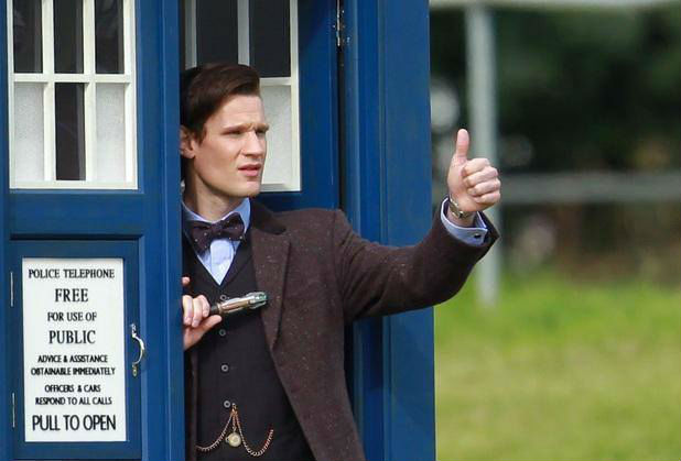
We've only got four exams, the longest of which being Physics at two and a half hours, but luckily they're spread out over four days, so I don't have to cram for two subjects at once beforehand. We've got two exams for Musculoskeletal Anatomy, one for Imaging Technique and one for Physics. I'm feeling confident for the Anatomy exam, as this is the one that, for now, makes the most sense. Physics on the other hand (see above image), well... writing pages and pages of notes has helped, but as far as 12-pulse bridge circuitry and rectifiers are concerned, I'm done for! You cannae change the laws of physics Jim, unfortunately...

Thankfully, a good chunk of some exams are MCQs (Multiple Choice Questions), and as long as you've done some reading around and understood the basics of the module, you should get most of these right, which usually can get you to the 40% pass boundary. Following those are the long answer questions, which range from 10-20 marks. 10 mark questions apparently only need about a page of writing, and 20 is two pages (...Wha?) So, my aim is to write as much as possible (within the relevant parameters anyway), and hope for the best!

After the exams are done with, we're back off out on placement again, but this time it'll be for 11 weeks! How will I survive 11 weeks of no Taekwon-do, nor a constant internet connection?! Do I risk taking my PS3, or shall I take boxsets upon boxsets of DVDs? Oh, decisions, decisions...

Wednesday, 9 April 2014

Portfolio Pains

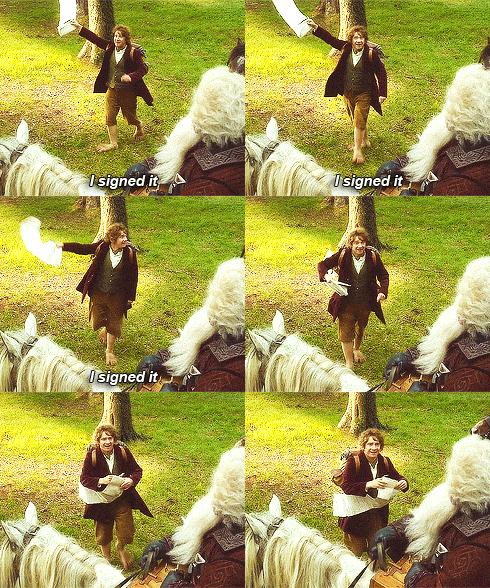
What it feels like when you finish an examination for your portfolio and you try to get the radiographer to sign it.

Stardate: 91875.79

So, I'm in my last week of placement for this block, and this week is my second one in clinical skills. This is basically a week where you can take on real examinations, just a little slower, and focus on assessment techniques, as you are with a lecturer practitioner, and not on a busy department. It's actually a really good opportunity to get examinations signed off for your portfolio, and brush up on what you know, which catches up on you really quickly.

It also allows you to work with students from other placement sites and see the variations, as every Trust has their own Local Rules and protocols regarding examinations and safety etc. For example, where I'm placed, we may do different routine projections for an examination to another site, or technique may vary. Again, it all depends on where you're placed, and what the radiographers there have shown you.

The realisation has pretty much finally dawned on me that being a student radiographer is actually really hard! Balancing 7 weeks of holiday, placements, lectures, travel and expenses claims, revision, portfolios "social life", hobbies and sleeping into one academic year is not an easy task. In fact, it's knackering. Especially for students like me, who are crazy enough to have a part-time job too!

Getting portfolios filled in can be difficult for some sections, especially ours, as where I'm placed, certain types of examinations are few and far between, e.g. spines. Sure, you see plenty of chests, knees etc, but when a spine comes in, everyone wants to do it! Luckily, I'm almost done with signing off my portfolio, I just need a few more unaided examinations, then I'll take on the assessment run! (Better not leave it until the last minute.)

It's amazing how quickly a year goes by, as although it feels like September was ages ago, it also feels like it was yesterday I was standing in General X-Ray getting in everyone's way! We've got two weeks of holiday for Easter (I don't have a clue what I'm going to do for two completely free weeks!), then two weeks of lectures, an assessment week, and then all placement weeks until Summer!

I've got two more days to sign off as many things as I can, then I can relax for once (with revision sittings of course). If I can find the time, I may even go and watch the new 'Captain America: Winter Solider'!

Well, as I like to say... Geronimo and LLAP!

References
  1. The Hobbit, n.d. gif, viewed 9 April 2014,<http://31.media.tumblr.com/60b475d6d7c7ece10f1cda53b21de510/tumblr_ms3tfrXwoW1r9hlt2o8_250.gif>.

Saturday, 22 March 2014

A Week in Fluoroscopy

This is the equipment used in my placement hospital, the Siemens Artis Zee

Stardate: 91823.58

It's that time again: placement! And for week one, I was placed in Screening (AKA Fluoroscopy); I was supposed to be in A&E, but it was changed at the last minute. If you didn't already know what fluoroscopy is, it's basically an imaging technique that uses real-time moving images of the internal structures.

The equipment I got the chance to learn to use properly was the Siemens Artis Zee Multi-Purpose, and it's controls, although they look confusing, take very little time to get used to. It's used in various types of fluoroscopic procedures:
  • Root nerve blocks - an injection that is given close to a nerve as it leaves the spine; usually for sciatica pain.
  • Facet joint injections - an injection of local anaesthetic or steroid which can anaesthetise the facet joint to block pain. Facet joints are small joints between each segment of the spine.
  • Barium enema - an x-ray examination of the large intestine to help diagnose any existing problems affecting that area. The colon is filled with a contrast material containing barium, which is done by pouring the contrast into the rectum.
  • Barium swallow - an x-ray examination of the upper gastrointestinal tract, including the oesophagus, and sometimes the stomach. The patient is asked to drink a suspension of barium sulphate, and the x-ray images are taken during the swallow. This procedure is done in various positions: AP, oblique and lateral, for a 3D assessment.
  • Myelogram - an x-ray examination that uses an injected contrast material to view the fluid-filled space (subarachnoid space) between the spinal vertebrae. 
I witnessed these examinations, and there are more that are carried out on the department. I certainly got used to wearing lead rubber aprons, and quickly learnt to check the apron first, as instead of picking up the lighter 0.25mm apron, I picked up a heavy 0.35mm apron, which made me feel faint after a while! I also started using the thyroid shield, just to be on the safe side.

Despite what everyone first believes about barium enemas, you barely see (or smell) anything, and nearly all fluoroscopic procedures are painless for the patient (the barium mixture patients have to swallow has a pretty nasty after-taste, mind). 

Overall, it was nice to see what the department does, and what this technique involves. It certainly lets you know if you want to go into that sort of speciality, and it does actually seem quite interesting. You certainly see a good variety of patients, too. It's made me start seriously looking into what I want to do post-graduation (still definitely Forensic Radiography) as there are so many options with Radiography!

Until next time guys! Next week I'm in Clinical Skills, so we'll see how that goes.

LLAP!

References

  1. Siemens Artis Zee Multi-Purpose, n.d. photograph, viewed 22 March 2014 <http://usa.healthcare.siemens.com/siemens_hwem-hwem_ssxa_websites-context-root/wcm/idc/groups/public/@global/@imaging/@angio/documents/image/mdaw/mtc0/~edisp/artis_zee_multi_purpose-00068177/~renditions/artis_zee_multi_purpose-00068177~10.jpg>.

Tuesday, 11 March 2014

Mayhem in March

When people ask you about mock exams; applies to both preparation and execution!

Stardate: 91796.42

As you may have noticed, it's been quite a while since I last posted (and it has been ages, because I was on placement last time!) This is generally down to mock exams having been placed over the last two weeks. We had our exams in Musculoskeletal Anatomy, Medical Imaging Science, and Diagnostic Imaging Technique. 

I would like to say I was happy with my results, and I will, but I'm a picky human being with a funny little brain, and I know I definitely could have done better. So I'm happy, but not satisfied with them (to be fair, I haven't had one set of results back yet). I was surprised when I concluded that I found the physics exam the least terrifying!

They weren't so terrible in retrospect, but the only thing to remember about radiography exams is: write as much as you can that's relevant to the question! I lost marks for assuming I didn't need to write something down. If you've learnt it, write it down and talk about it! Also another handy tip is to definitely keep on top of your revision. It soon builds up if you don't!

Anyway, since I've managed to free myself from revision, as a 'reward', I've been reading some radiography journals to pass some time (when I'm not watching Star Trek, or Doctor Who). Two articles caught my eye in last month's (Feb 2014) issue of the journal Radiography. These were very different in content, but interesting reads.

The first was about using cross-sectional imaging in virtual post-mortem examinations. This appealed to me, generally because it's to do with Forensic Radiography, which is the field in which I want to continue my studies. The second was about professionals' experiences of imaging in the radiography process. I may have mentioned it before, but if you haven't already, signing up to the SoR is very beneficial for (prospective) radiography students, as you can access journals and articles, and loads of information which can give a nice scope of the field.

Soon you'll be hearing more from me again, as I'm back on placement next week! I'll be in A&E for one week, clinical skills for two, and a screening room for another. So there'll be plenty to get on with, and hopefully I'll have interesting things to feed back!

LLAP and reverse the polarity of the neutron flow!

References
  1. Matt Smith Filming His Final Scene, n.d. photograph, viewed 11 March 2014 <http://static.fjcdn.com/comments/Matt+Smith+after+filming+his+final+scene.+Leaving+the+TARDIS...for+_76a0889eed211250bf3e27e7c154b0c1.jpg>.

Wednesday, 22 January 2014

Working in Theatre and ORIF Surgery

A scan of a Weber's Category B fracture.

Stardate: 91665.06

Day three of my placement, and luckily once again I was in theatre, and I have to say I'm really enjoying it. I saw one spinal examination today on the lumbar spine, and an ORIF (Open Reduction and Internal Fixation) surgery on an ankle.

The spinal examination required little screening, and these were few and far between, which meant half of the time was spent not being able to see anything and standing around waiting to be needed. 

This led me to think of what a student radiographer needs to know before going into theatre. My general thoughts were just tips on how to feel less overwhelmed by theatre situations, as it is a whole new environment:
  • know the theatre rules and the theatre culture
  • make sure you're wearing the right personal protection equipment - e.g. hat, mask, lead-rubber aprons
  • once the patient's skin is open, assume everything is sterile - don't touch anything except the radiography equipment and you'll be okay!
  • know how to use the image intensifier (C-arm - a portable C-shaped x-ray device used for imaging during surgical procedures), or observe it being used before undertaking it yourself
  • know what is needed during the procedure, if unsure, ask your supervisor
  • go to the toilet before surgery - you can be in there for a really long time!
  • and if you feel faint, back up to the wall and slide to the floor

The second case was one I attended almost fully unassisted by my supervisor. I felt confident enough to handle the C-arm by myself, and the only hindrance I faced was the positioning of the patient on the table, as they were a little lower down than normal, which meant a bit of the table's metal framework was unavoidably in the image, but not obscuring it. This case was the ORIF ankle case. Open Reduction is surgery to set bones, and Internal Fixation is the fixing of screws and/or plates to facilitate healing. This surgery is used when casting or splinting a fracture alone would not assist repair.

This went well and I was quite impressed at my being pro-active with getting the imaging done and feeling that I could do the examination. The patient had a Weber's fracture (ankle fracture) on the medial malleolus (inner side) of the ankle, and the plates were being placed so the bone was able to heal correctly. I was given positive feedback, and I think I'm much more confident than I was before on placement, and I'm starting to get into the hang of working within different departments.

I think this has been my favourite week so far! Now to do a quick workout, eat a cookie and watch 'An Idiot Abroad' in bed!

LLAP!

References
  1. Jeremy Jones, Radiopedia.org, photograph, viewed 22 January 2014 <http://images.radiopaedia.org/images/341127/186015a2d8111ae7f0232b0fb61ab9.jpg>.

Tuesday, 21 January 2014

Surprise Birthday Spondylosyndesis!

An example of a post-operative multi-level cervical fusion.

Stardate: 91662.61

Overall, I have to say, today wasn't a bad birthday. I wasn't knackered upon arriving at the hospital, I'd had a decent breakfast, and I got to spend pretty much all of my day in orthopaedic theatre! I saw three cases, all of them spinal surgery, and it was great! I saw two lumbar, and a cervical spine case, and the latter was just amazing to witness. (See, I told you my posts would hopefully get more interesting!)

The patient had been suffering with a cervical spinal disc herniation between the C4 and C5 vertebrae. This is basically where a tear in the outer fibrous layer (annulus fibrosus) of the inter-vertebral disc has allowed the central jelly-like portion (nucleus pulposus) to bulge out and cause mechanical compression (pressure) upon a nerve (in this case, the spinal cord). In order to relieve this patient of their pain, surgery was undertaken, and in this case it was a spinal fusion (or spondylosyndesis). 

The type of fusion used for this particular case was ACDF (Anterior Cervical Disectomy and Fusion). This is a surgical procedure where the surgeon enters the space between the two discs through a small incision anteriorly (from the front) and to either the right or left side of the neck. The affected disc is then completely removed, and the intervertebral foramen (the channel through which the spinal cord runs) is then enlarged by a drill to allow the nerve more room. A device is placed between the two vertebrae to maintain spine alignment, and is then fixed with a 'cage' using metallic pedicle screws made from titanium. This type of surgery can take 5 hours, and although I was only in for a few of them, I saw quite a lot.

The consultant surgeon was incredibly nice and explained everything as he went along, and why he was doing it. Before we properly started he showed me some of the patient's previous MRI scans, which helped me to understand what we were actually seeing and scanning. When he'd finished doing different stages, he even encouraged me to come over and have a good look at his work, and I felt quite privileged by it, as some of the surgeons I've seen are quite offish and uninterested.

When I wasn't in theatre, I was out on mobiles, so I got to visit the ICU (Intensive Care Unit) and Oncology. I finally put into practice what I'd learnt about moving and handling (how to move yourself and patients safely), as many patients on these wards find it difficult to get into position for their x-rays. One of the elder male Oncology patients made me smile, as he was fascinated by the mobile x-ray tube, as he thought we were coming to take him back to Main X-Ray (which is about a mile away from Oncology), and he asked quite a lot of questions, and praised the technology's speed.

To finalise this post, I really had a great day in theatre, and I'm looking forward to the possibility of going tomorrow! And, I didn't faint! I knew I wouldn't!

I'm off to spend the remainder of my birthday relaxing in bed with my Spider-Man pillow, some bicuits and 'An Idiot Abroad' DVDs...

LLAP!

References
  1. Alexios Apazidis, spineuniverse.com, photograph, viewed 21 January 2014 <http://cloud2.spineuniverse.com/sites/default/files/imagecache/inline-content/images/2012/07/25/4-months-postoperative.jpg>

Monday, 20 January 2014

Return of the Placement!

As insane as it sounds, it's slightly true.

Stardate: 91659.59

So, I'm back out on placement already, and my first day started off in a bit of a panic. Why? Well, because I thought it was okay to not check the TLD (Thermoluminescent dosimetry) I was handed before getting the train to placement. As a result, I found out I had the wrong TLD, which brought forward the looming possibility of not being able to go on placement until I had the correct one, which could take up to 3 days...

Luckily, the Radiation Supervisor and my clinical tutor said that one not having a TLD for a couple of days wouldn't be a major risk... unless something drastic happened. So I decided to risk it and get on placement anyway.

I was glad that I did. I managed to get lots of examinations for my portfolio signed off, mainly chest x-rays, because there weren't many portable examinations being requested. I saw some interesting departments like Oncology and ICU, and was glad to find out I hadn't forgotten everything since last year, which I consider to be a bonus. There were one or two situations that happened involving fainting patients and the like, but it was all good experience, and you learn from them quickly. Being in the department with other students can be helpful as looking at each others x-rays is fun, and good practice, as you get to see some pretty amazing things, and wince in unison at some painful ones.

I'm hoping that tomorrow I get to go into theatre, because tomorrow happens to be my birthday, and I want it to be interesting. I don't tend to make a big deal about them, but being the strange person I am, I think it'd be great fun to get some time in theatre to learn how the techniques are adapted to different cases. And who doesn't like a bit of gore now and then?

As for spare time, in between what little sleep I tend to get and now being out on clinical, I've started doing an online Forensic Science course, which entails studying the field using a 'Murder on the Loch' case study, while I sit happily listening to Mozart. It's fascinating, and as it's linked to what I want to do in the future, some of the material slots in nicely. It seems one has gone back into her geeky-recluse mode once more...

LLAP and the game is now most definitely on.

References
  1. Sherlock and Violins, n.d. photograph, viewed 20 January 2012 <https://i.chzbgr.com/maxW500/6564377344/hBA076E54/>